Accessory Navicular Syndrome (ANS)
Quick Facts
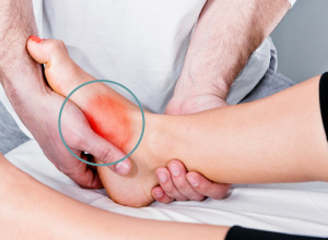
- Accessory navicular syndrome (ANS) happens when an abnormal foot growth called the navicular bone (os navicularum or os tibiale externum) creates pain on the side of the foot between the medial arch and heel.
- Though having extra bones in the foot is a common orthopaedic condition, only a very small portion of the population (between 2-14%) has an accessory navicular bone (located within the posterial tibial tendon), and an even smaller percentage of that group experiences pain from the uncommon bone growth.
- Because accessory navicular syndrome rarely causes symptoms, treatment is often unnecessary. When pain occurs, treatment usually includes ice, physical therapy and orthotics.
Symptoms Of Accessory Navicular Syndrome
The presence of an accessory navicular bone is congential (meaning it is usually present at birth) and rarely causes any symptoms. In fact, most people aren’t aware they have the extra piece of bone or cartilage until it shows up on an X-ray taken for another problem.
In rare cases, the accessory navicular bone creates a bony prominence in the midfoot that causes pain, redness and swelling in the medial arch area, plantar fasciitis, bunions and heel spurs. When this happens, the condition is called accessory navicular syndrome. ANS can arise from a number of things, including foot trauma like ankle sprains and twists, overuse, excessive physical activity, and irritation from footwear. These symptoms generally present themselves during adolescence, when the cartilage in the foot is still maturing into bone. But ANS can also become symptomatic in adulthood.
Common Treatment For ANS
Most of the time, the presence of an accessory navicular bone is asymptomatic and the harmless condition usually doesn’t require treatment. There is no need for intervention if the small “accessory” doesn’t cause pain.
On the rare occasion that the accessory navicular bone creates symptoms, there are a number of treatment options available to help ease the pain and discomfort. Like with many foot ailments, these include icing the area, taking over-the-counter pain medications, immobilization, physical therapy and wearing either orthotics, a cast or a removable walking boot.
Physical therapy is the often prescribed method of treatment, as it is a non-invasive way to strengthen the surrounding muscles and relieve symptoms. Specially designed orthosis follow, as they can help prevent future symptoms from presenting themselves.
Some patients turn to prolotherapy, which is an injection technique that can help strengthen the ligament, tendon and muscle attachments in the problem area by causing an anti-inflammatory response in the tissue. The injections cause the body to send blood, regenerative cells and collagen cells to the area, which can help the tissue become stronger.
Only rarely is surgical removal of the accessory navicular bone advised. When this happens, the orthopaedic surgeon will remove the accessory bone and begin foot reconstruction by repairing the posterior tibial tendon.
Notice concerning medical entries:
Articles having medical content shall serve exclusively for the purpose of general information. Such articles are not suitable for any (self-) diagnosis and treatment of individual illnesses and medical indications. In particular, they cannot substitute for the examination, advice, or treatment by a licensed physician or pharmacist. No replies to any individual questions shall be effected through the articles.


